


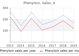
Phenytoin
"Generic phenytoin 100 mg, medications causing hair loss."
By: Paul Reynolds, PharmD, BCPS
- Critical Care Pharmacy Specialist, University of Colorado Hospital
- Clinical Assistant Professor, Department of Clinical Pharmacy, Skaggs School of Pharmacy and Pharmaceutical Sciences, University of Colorado, Aurora, Colorado

http://www.ucdenver.edu/academics/colleges/pharmacy/Departments/ClinicalPharmacy/DOCPFaculty/Q-Z/Pages/Paul-Reynolds,-PharmD.aspx
Retnal laser treatment depends on the far more gentlemanly tssue efect known as photocoagulaton symptoms glaucoma buy 100 mg phenytoin visa. That induces an elevaton in the temperature of the tssue that spreads out from the pigment and literally cooks the surrounding tssue at a microscopic level treatment 7 order phenytoin with a visa. The resultng coagulaton of proteins causes the desired efect? hopefully without any photoablatve or photodisruptve pyrotechnics medicine express discount phenytoin 100 mg without prescription. In this case symptoms miscarriage generic phenytoin 100 mg overnight delivery, a very low-power laser is used to actvate a specifc chemical to obtain the desired efect in the tssue. The use of a red laser to actvate verteporfn (Visudyne) in order to treat neovascular age-related macular degeneraton is the best example of this. Figure 1 is the classic display of how each laser color is absorbed in various ocular tssues. For a long tme, people hoped that diferent colors would allow one to customize the treatment depending on the indicaton. Perhaps more clinically signifcant is both the marked drop-of in hemoglobin absorpton and the gradual drop-of in melanin uptake as you move into the red end of the spectrum. This explains in part why red and infrared burns require more power and tend to penetrate deeper into the more pigmented choroid. It also helps explain why the infrared diode laser in partcular is so diferent to use relatve to a green laser. Note that, in general, the further you go toward red, the less the absorpton?hence the need for more power and a resultant deeper burn with longer wavelengths. You can also see how yellow hits a peak of oxyhemoglobin absorpton relatve to green, which accounts for the diference in how microaneurysms are afected by each wavelength. Finally, you can see why it is a very bad idea to use blue light anywhere near the fovea, where xanthophyll pigment is found. Besides, you will basically be using whatever laser has been plopped in your clinic because there is no way you can go out and shop and compare with these enormously expensive devices. Fortunately, most of the studies on diabetc retnopathy were performed using some sort of green wavelength?usually argon green or its kissin? cousin diode green?and that is prety much the standard color of laser found anywhere. Modern solid state lasers can generate green, yellow and red wavelengths, and some retnal specialists enjoy using a yellow laser because it seems to allow more selectve treatment of retnovascular lesions and they feel there is less long term scar formaton. A red laser can be useful when there are media opacites because the longer wavelength can get through beter than green. An infrared laser can penetrate even beter, but that wavelength is so diferent to use that it gets its own chapter at the end of the book. However, no large study has proven that there is a defnite diference between wavelengths in terms of clinical outcome. In fact, the Diabetc Retnopathy Clinical Research Network allows interchangeable use of yellow and green lasers for focal treatment of macular edema. For simplicity, the rest of this book will assume you have some sort of green laser to work with because that is the most common. The one thing to remember with any wavelength is what you learned in second-grade science class: Black absorbs everything and white refects everything. In other words, if you are treatng happily in an area of the retna and you come upon a dark area like a nevus or a previous laser scar, you need to watch out, because you can get an explosive burn as the pigment sucks in the laser (higher absorpton translates into higher photothermal elevaton). Remember to Turn It Down When You Hit Brown (and Cut Way Back When You Hit Black). Alternatvely, if you need to treat a very pale area, you will need to crank it up?but be super careful when you hit pigment again. Here, for completeness, are the only formulas in the book: Irradiance (W/cm2) = Power (Watts) Spot Area (cm2) Energy (Joules) = Power (Watts) x Time (Seconds) Fluence (J/cm2) = Power (Watts) x Time (Seconds) Energy (Joules) 2 = 2 Spot Area (cm) Spot Area (cm) We will try to stay away from the obligatory discussion of energy, work, radiometric terminology, etc. The key thing is that your laser output has a certain level of mojo and you need to know exactly how to control it. Note that going up or down on power (Wats) or on exposure duraton (Time) creates a linear increase or decrease in the energy delivered. This means that if you are getng a good burn and you decide to, say, double the exposure duraton, then you have to decrease the power or you will really cook things. It is hard to imagine why on earth one would want to do this when one is getng a good burn, but this is always mentoned in basic laser texts and it does help ensure that you understand the relatonship. The really signifcant thing is that the clinical efect tends to be very intuitve?a mild increase in the power or duraton will give you a mild increase in your burn, and the same is true if you want to turn things down. However, because we are dealing with a biological system and not a photometer, it turns out that the relatonship between the energy delivered and the type of burn that you get is a bit more complex. The exact same energy can result in diferent burns because the burn depends on how the laser is absorbed and how the heat is transmited by the tssue. In other words, fddling with the laser power and duraton generally results in a common-sense change in the degree of uptake?a litle more tme or power results in a litle more burn and a lot more tme or power results in a lot more burn. Colorful Box Break: Since you are basically using your laser to warm up the retna, you do need to be careful about using high powers at short duraton, because the nice linear relatonship breaks down and you can end up microwaving the proverbial poodle of urban legend. Unfortunately, heat transfer is governed by factors far more complicated than the weenie-pre-med-physics equaton above. For instance, heat transfer explains why it is easy to get a burn in the retna but really hard to get a burn on a big blood vessel?the blood carries? away the heat and you can?t get the vessel wall to cook easily. Because you should treat a big vessel exactly never, just remember that if you use a lot of power over a really short duraton, there isn?t tme for the heat to spread out and you can get a much hoter burn than you would expect if the response of the tssue were truly linear. Modern lasers, and especially the patern lasers discussed below, use very short duratons, and it is felt that those shorter duratons allow careful ttraton of how far the burn spreads. But you need to internalize the fact that the tssue response to your laser is nowhere near as linear as the fuence equaton might suggest. In other words, if you were getng a good burn using 100 milliwats for 100 milliseconds, and if you decided to decrease the duraton by a factor of 10 to 10 milliseconds, the fuence equaton suggests that you will need to turn the power up by that same factor of 10 to 1000 milliwats to get the same burn. But you would never, ever really do that?you would vaporize the retna because the rapid buildup of heat would not have tme to dissipate. This is very important to grasp; a small change in spot size can make a big diference in the energy you pour into the retna if you don?t compensate by changing the power or duraton. This is especially likely if you are also using a brief duraton (less tme for heat transfer, remember). The result is an explosively expanding bubble of water vapor that will cause a hole or hemorrhage or both. You can really mess up an eye doing this? and lose lots of style points with your patents and colleagues. We will return to this concept several tmes in this book to be sure it sinks in?it has to be internalized to your lizard brain parts just like the mental switch that keeps you from engaging phaco when you are next to the posterior capsule. Anyway, if you make the spot size smaller?even if it is only a litle bit smaller?you really have to be religious about decreasing the other parameters so that you don?t start punching holes in the retna. For instance, you might be using a strong power to cut through media opacites and you might also be using a short duraton to try to make the laser less painful for the patent (no worries?much more on these techniques later). You might then decide to decrease the spot size in order to get an even beter burn?a smaller spot will not spread out as much as a large spot if the view is hazy. If you do this, then you must cut back on power and work your way back up to a safe burn; otherwise, you will have increased irradiance and fuence by the square of the diference in spot size, and you will very likely cause a dangerously hot burn. Repeat: You must cut back on power and work your way back up to a safe burn if you decrease the spot size. As we will see in the next chapter, each type of contact lens will minify or magnify the size of the actual spot projected on the retna. If you switch to a diferent contact lens, you might be shrinking the actual spot size without realizing it? thus dramatcally changing how much power is focused onto the retna. Or if you are doing a macular laser in an area of swollen retna, the thickened retna will tend to difuse the beam, and when you move *Repeat: this concept needs to be so ingrained that if you are captured by aliens and pithed for a science project your decerebrate hands will stll reach for the power knob if someone says smaller spot. Or when you are startng your laser career, it may take way longer than you want to get anything into focus and you may decide to fre away before your focus is crisp because you are frustrated. If the gods of retna then suddenly put your aiming beam into perfect focus, your spot will shrink down and suddenly you will be burning holes in important parts of your patent. This will all be covered in greater detail in upcoming chapters?but the point is that your spot size may change whether you want it to or not, and you have to be ready to antcipate these changes and alter your parameters accordingly. There is yet another way that the biology of lasering can get you into trouble even without using small spots, and this occurs when you are using powers, for whatever reason, that are causing very hot burns. Heat building up in the periphery of the burn can at least dissipate into untreated retna, but heat building up in the center of the burn is trapped and cannot spread out much.
Some authorities advocate that the amount of solid food eaten should not exceed the amount that patients would ordinarily be eating at their target weight symptoms precede an illness buy phenytoin 100 mg cheap. Expanding cuisine options is important to medications with sulfa buy phenytoin us avoid the severely restricted food choices fre Treatment of Patients With Eating Disorders 41 Copyright 2010 340b medications discount phenytoin 100 mg without prescription, American Psychiatric Association medications canada best order for phenytoin. Legitimate food allergies and patients? religious and cul tural practices must be considered and discussed to limit patient rationalizations for restricted eating. Intake levels should usually start at 30?40 kcal/kg per day (approximately 1,000?1,600 kcal/day). During the weight gain phase, intake may have to be advanced progressively to as high as 70?100 kcal/kg per day for some patients; many male patients require a very large num ber of calories to gain weight. Patients who require significantly higher caloric intakes may be discarding food, vomiting, or exercising frequently or they may engage in more nonexercise motor activity such as fidgeting; others may have a truly elevated metabolic rate. Patients re quiring much lower caloric intakes or those suspected of artificially increasing their weight by fluid loading should be weighed in the morning after voiding while they are wearing only a gown; their fluid intake also should be carefully monitored. Assessing urine specimens obtained at the time of weigh-in for specific gravity may help ascertain the extent to which the measured weight reflects excessive water intake. Particularly in residential or hospital treatment programs, it may initially be difficult to ob tain the cooperation of patients who do not wish to be there. In addition, many patients have delayed gastric emptying that initially impairs their ability to tolerate 1,000 calories/day. During hospitalization, giving patients a liquid feeding formula in the early stages of weight gain and then gradually exposing them to food and slowly increasing their activity level can be a very effective strategy for inducing weight gain (114). As patients are able and as their cooperation improves, a 2?3 lb/week gain in residential or hospital programs can be ex pected without compromising the patients? safety. In addition to an increased caloric intake, patients also benefit from vitamin and mineral supplements. Serum potassium levels should be regularly monitored in patients who are per sistent vomiters. Hypokalemia should be treated with oral or intravenous potassium supple mentation and rehydration. Physical activity should be adapted to the food intake and energy expenditure of the patient, taking into account bone mineral density and cardiac function. For the severely underweight patient, exercise should be restricted and always carefully supervised and monitored. Once a safe weight is achieved, the focus of an exercise program should be on physical fitness as op posed to expending calories. The focus on fitness should be balanced with restoring patients? positive relationship with their bodies?helping them to take back control and get pleasure from physical activities rather than being compulsively enslaved to them. An exercise program should involve exercises that are not solitary, are enjoyable, and have endpoints that are not de termined by time spent expending calories or changing weight and shape. Staff should help patients deal with their concerns about weight gain and body image changes, given that these are particularly difficult adjustments for patients to make. In fact, there is general agreement among clinicians that distorted attitudes about weight and shape are the least likely to change and that excessive and compulsive exercise may be one of the last of the behaviors associated with an eating disorder to abate. Although it is by no means certain that patients? abnormal eating habits will improve simply as a function of weight gain (116), there is considerable evidence to suggest that other eating disorder symptoms diminish as weight is restored with nutritional rehabilitation. For example, clinical experience indicates that with weight restoration, food choices increase, food hoarding decreases, and obsessions about food decrease in frequency and intensity, although they do not necessarily disappear. Providing anorexia nervosa patients who have associated binge eating and purging behaviors with regular structured meal plans may also enable them to improve. For some patients, how ever, giving up severe dietary restrictions and restraints appears to increase binge-eating behav ior, which is often accompanied by compensatory purging. As weight is regained, changes in associated mood and anxiety symptoms as well as in phys ical status can be expected (117). Clinicians should advise patients of what changes they can anticipate as they start to regain weight. In the initial stages, the apathy and lethargy associated with malnourishment may abate. However, as patients start to recover and feel their bodies be coming larger, and especially as they approach frightening magical numbers on the scale that represent phobic weights, they may experience a resurgence of anxious and depressive symp toms, irritability, and sometimes suicidal thoughts. These mood symptoms, non-food-related obsessional thoughts, and compulsive behaviors, although often not eradicated, usually decrease with sustained weight gain. Weight gains result in improvement in most of the physiological complications of semi starvation, including improvement in electrolyte levels, heart and kidney function, and atten tion and concentration. Initial refeeding may be associated with mild transient fluid retention, and patients who abruptly stop taking laxatives or diuretics may experience marked rebound fluid retention for several weeks, presumably from salt and water retention caused by elevated aldosterone levels associated with chronic dehydration. Patients may experience abdominal pain and bloating with meals from the delayed gastric emptying that accompanies malnutrition. Constipation, which may be ameliorated with stool softeners, can progress to obstipation and, rarely, acute bowel obstruction. As weight gain pro gresses, many patients also develop acne and breast tenderness. Many patients become unhappy and demoralized about resulting changes in body shape. Management strategies for dealing with these milder adverse effects include careful refeeding, frequent physical examinations, and forewarnings to patients about mild refeeding edema. A severe refeeding syndrome may occur when severely malnourished patients (generally those weighing <70% of their healthy body weight) are re-fed too rapidly, particularly in the context of enteral or parenteral feedings but also with vigorous oral refeeding regimens. This syndrome consists of hypophosphatemia, hypomagnesemia, hypocalcemia, and fluid reten tion. In some case series, the refeeding syndrome has been reported to occur in roughly 6% of hospitalized adolescents (118). Excessively rapid refeeding and nasogastric or parenteral feeding may be particularly dan gerous because of their potential for inducing severe fluid retention, cardiac arrhythmias, car diac failure, respiratory insufficiency, delirium, seizures, rhabdomyolysis, red cell dysfunction, and even sudden death, especially in the lowest-weight patients (118, 119). In such cases, phos phorus, magnesium, and/or potassium supplementation will be necessary (118, 120). In one se ries of hospitalized adolescents, moderate hypophosphatemia occurred in 5. Besides monitoring of mineral and electrolyte levels, general medical monitoring during re feeding should include assessment of vital signs, monitoring of food and fluid intake and out put (if indicated), and observation for edema, rapid weight gain (associated primarily with fluid overload), congestive heart failure, and gastrointestinal symptoms. For children and adolescents who are severely malnourished (weigh <70% of their standard body weight), cardiac monitor ing, especially at night, may be advisable (120). Some patients are completely unable to recognize their illness, accept the need for treatment, or tolerate the guilt that would accompany eating, even when performed to sustain their lives. On the rare occasions when staff have to take over the responsibilities for providing life preserving care, nasogastric feedings are preferable to intravenous feedings. In some programs, supplemental overnight pediatric nasogastric tube feedings have been used to facilitate weight gain in cooperative patients. This practice is not routinely recommended at present, although it appears to be well tolerated, may slightly decrease hospital stays in children, and may be ex perienced positively by some patients, particularly younger patients, who may feel relieved to Treatment of Patients With Eating Disorders 43 Copyright 2010, American Psychiatric Association. If used, such interventions should never supplant expectations that the patient will resume normal eating patterns on his or her own. Total paren teral feeding is required only rarely and for brief periods in life-threatening situations. Forced nasogastric or parenteral feeding can each be accompanied by substantial dangers. When nasogastric feeding is necessary, clinical experience suggests that continuous feeding. As an alter native to nasogastric feedings, in very difficult situations where patients physically resist and constantly remove their nasogastric tubes, gastrostomy or jejunostomy tubes may be surgically inserted. As described above, rapid refeeding can be associated with the severe refeeding syn drome, and infection is always a risk with parenteral feedings in emaciated and potentially im munocompromised patients with anorexia nervosa. Consequently, these interventions should not be used routinely but should be considered only when patients are unwilling or unable to coop erate with oral feedings or when the patients? health, physical safety, and recovery are being threatened. If using interventions that patients with anorexia nervosa may experience as coercive, the clinician should consider the potential impact on the therapeutic relationship, especially since maintaining a sense of control is often a key dynamic in these patients. During the last few years, there has been considerable debate about the ethics of involun tarily feeding patients with anorexia nervosa (122, 123). There is general agreement that chil dren and adolescents who are severely malnourished and in grave medical danger should be re fed, involuntarily if necessary, but that every effort should be made to gain their cooperation as cognitive function improves. Ethical as well as clinical dilemmas often confront clinicians dealing with adult patients with chronic anorexia nervosa and their families. The general principles to be followed are those di recting good, humane care; respecting the wishes of competent patients; and intervening re spectfully with patients whose judgment is severely impaired by their psychiatric disorders when such interventions are likely to have beneficial results (124, 125).
Buy cheapest phenytoin. Kirsty shares her optic neuritis MS symptom story 2 of 2.
Subserous intensity focused ultrasound treatment lice phenytoin 100 mg sale, myolysis/radiofrequency myomas must be distinguished from ovarian tumors treatment thesaurus cheap 100mg phenytoin amex. Leio? ablation 911 treatment center purchase cheap phenytoin online, and laparoscopic or vaginal occlusion of uterine myosarcoma is an unusual tumor occurring in 0 symptoms jaw cancer order phenytoin on line amex. Emergency Measures tility, myomectomy can be offered, but patients should be Emergency surgery may be required for acute torsion of a counseled that recurrence is common, postoperative pelvic pedunculated myoma. If the patient is markedly anemic as a adhesions may impact fertility, and cesarean delivery may result of long, heavy menstrual periods, preoperative treat? be necessary. The only emergency indication for myomectomy during pregnancy is For acute abdomen associated with an infarcted leiomyoma torsion. J Clin Endocrinol hyperplasia almost completely with the use of oral contra? Metab. Prevalence, symptoms and management Staging and prognosis are based onsurgical and pathologic of uterine fbroids: an international internet-based survey of evaluation only. Treatment Treatment consists of total hysterectomy and bilateral sal? pingo-oophorectomy. Abnormal bleeding is the presenting sign in 90% examination is routinely taken and lymph node sampling of cases. After a negative pregnancy test, endometrial postoperative irradiation is indicated. It occurs most advanced or metastatic endometrial adenocarcinoma may often in women 50-70 years of age. Obesity, nulliparity, be accomplished with large doses of progestins, eg, diabetes, and polycystic ovaries with prolonged anovula? medroxyprogesterone, 400 mg weekly intramuscularly, or tion, unopposed estrogen therapy, and the extended use of megestrol acetate, 80-160 mg daily orally. Prognosis (hereditary nonpolyosis colorectal cancer, Lynch syn? drome) are at significantly increased risk, with a lifetime With early diagnosis andtreatment, the overall 5-year sur? incidence as high as 30%. With stage I disease, the depth of myome? Abnormal bleeding is the presenting sign in 90% of trial invasion is the strongest predictor of survival, with a cases. Any postmenopausal bleeding requires investiga? 98% 5-year survival with less than 66% depth of invasion tion. Pain generally occurs late in the disease, with metas? and 78% survival with 66% or more invasion. When to Refer atypical endometrial cells but are an insensitive diagnostic All patientswith endometrial carcinoma should be referred tool. Simultaneous hysteroscopy can be a valuable addition in order to localize polys or other lesions within the uterine cavity. History of prolonged vulvar irritation, with pruritus, response, fuorinated steroids should be replaced with local discomfort, or slight bloody discharge. Early lesions may suggest or include non-neoplastic lichen sclerosus, recommended treatment is clobetasol epithelial disorders. Late lesions appear asa mass, anexophytic growth, once daily until symptoms resolve. As with squamous celllesions of the cervix, a uninvolved portions of the vulva may be spared. To avoid the morbidity of inguinal lymphadenectomy, some guidelines recommend sentinel Benign vulvar disorders that must be excluded in the lymph node sampling for women with early-stage vulvar diagnosis of carcinoma of the vulva include chronic cancer. Patients with more advanced disease may receive granulomatous lesions (eg, lymphogranuloma venereum, preoperative radiation, chemotherapy, or both. Lichen sclerosus and other associated leukoplakic changes in the skin should be biopsied. Prognosis superimposed vulvar cancer will develop in a woman Basal cell vulvar carcinomas very seldom metastasize, and with a nonneoplastic epithelial disorder (vulvar dystrophy) carcinoma in situ by definition has not metastasized. Diagnosis less, without inguinal lymph node metastases, have an 85-90% 5-year survival rate. Multiple skin? punch specimens can be taken in the office under local anesthesia, with care to include tissue from the edges of. Colposcopy of vulva, vagina, and All patients with invasive vulvar carcinoma should be cervix can help in identifing areas for biopsy and in plan? referred to a gynecologic oncologist. The role of preoperative ultrasound evalua? Vulvar cancer generally spreads by direct extension into tion of inguinal lymph nodes in patients with vulvar malig? the vagina, urethra, perineum, and anus, with discontinu? nancy. Diagnosis and treatment options of vulvar required except in advanced cases for planning therapeu? cancer: a review. The optimum duration of therapy is not clear, and the relative merits in terms of side effects and long-term risks and benefts show insignifcant differences when compared with each other and, in mild? General Considerations Any of the combination oral contraceptives, the con? Endometriosis is an aberrant growth of endometrium out? traceptive patch, or vaginal ring may be used continu? side the uterus, particularly in the dependent parts of the ously for 6-12 months. Breakthrough bleeding can be pelvis and in the ovaries, whose principal manifestations treated with conjugated estrogens, 1. Progestins, specifically oral norethindrone acetate and esis and natural course are not fully understood. Clinical Findings However, they are not superior to other methods such as combined oral contraceptives as frst-line therapy. Side effects of vasomotor number of women with endometriosis, however, remain symptoms and bone demineralization may be relieved asymptomatic and most women with endometriosis have a by "add-back" therapy, such as conjugated equine estro? normal pelvic examination. Danazol is an androgenic drug that has been used for decreased uterine mobility, cervical motion tenderness, or the treatment of endometriosis-associated pain. However, danazol has a high inci? endometrial tissue may produce blood in the stool that dence of androgenic side effects that are more severe must be distinguished from bowel neoplasm. Ultimately, a definitive diagnosis of endometriosis is or letrozole) has been evaluated in women with chronic made only by histology of lesions removed at surgery. Laparoscopic ablation of endometrial is effective in the amelioration of pain associated with implants significantly reduces pain. However, there is no evidence that any of and, if necessary, removal of ovarian endometriomas these agents increase the likelihood of pregnancy. Their enhance fertility, although subsequent pregnancy rates are preoperative use is of questionable value in reducing the inversely related to the severity of disease. In premeno? tors may include vaginal birth, genetic predisposition, pausal women, hormone replacement then may be used to advancing age, prior pelvic surgery, connective tissue dis? relieve vasomotor symptoms. However, hormone replace? orders, and increased intra-abdominal pressure associated ment may lead to a recurrence of endometriosis and asso? with obesity or straining associated with chronic constipa? ciated pain. Clinical Findings There is little systematic research regarding either the pro? Symptoms ofpelvic organ prolapse may include sensation gression of the disease or the prediction of clinical out? of a bulge or protrusion in the vagina, urinary or fecal comes. The prognosis for reproductive function in early or incontinence, constipation, a sense of incomplete bladder moderately advanced endometriosis appears to be good emptying, and dyspareunia. Hysterectomy, with bilateral lapse, including prolapse of the uterus, vaginal apex, and salpingo-oophorectomy, often is regarded as definitive anterior or posterior vaginal walls, is likely multifactorial. Treatment symptoms may recur in women even after hysterectomy the type of therapy depends on the extent of prolapse and and oophorectomy. Supportive measures include a high-fber diet and laxatives to improve constipation. Rarely necessary except for acute abdomen associated with Pelvic muscle training (Kegel exercises) is a simple, nonin? ruptured or bleeding endometrioma. Pathogenesis and pathophysiology of endome? the most common surgical procedure is vaginal or abdom? triosis. Consensus on current management of endometrio? pension by either uterosacral or sacrospinous fixation vagi? sis. Advances in pharmacotherapy for treating endome? dures, an anti-incontinence procedure should be consid? triosis. If the patient desires pregnancy, the same proce? dures for vaginal suspension can be performed without. General Considerations hysterectomy, though limited data on pregnancy outcomes or prolapse outcomes are available. For elderlywomen who Cystocele, rectocele, and enterocele are vaginal hernias do not desire coitus, colpocleisis, the partial obliteration of commonly seen in multiparous women. Uterine sus? hernia of the bladder wall into the vagina, causing a soft pension with sacrospinous cervicocolpopexy may be an anterior fullness. Cystocele may be accompanied by ure? effective approach in older women who wish to avoid hys? throcele, which is not a hernia but a sagging of the urethra terectomy but preserve coital function. When to Refer into the posterior vagina, causing a collapsible pouch-like fullness. Enterocele is a vaginal vault hernia containing Refer to urogynecologist or gynecologist for inconti? small intestine, usually in the posterior vagina and result? nence evaluation. Female Pelvic Med No single historical, physical or laboratory finding is Reconstr Surg.
The urinary porphyrins and several haeme enzymes can be used as early biomarkers of arsenic toxicity (Garcia-Vargas & Hernandez-Zavala medicine hollywood undead order 100mg phenytoin otc, 1996) treatment nurse order 100mg phenytoin free shipping. Rodents exposed for 6 weeks to 5 asa medications purchase phenytoin 100mg on-line sodium arsenate in drinking-water showed a substantial increase in the urinary excretion of porphyrins treatment quality assurance unit purchase phenytoin 100mg line, with excretion of uroporphyrin exceeding that of coproporphyrin (Woods & Fowler, 1978). Livers of animals were homogenized and mitochondria and microsomal fractions were then prepared. Continuous prolonged expo sure to sodium arsenate resulted in depression of hepatic? Concomitantly, urinary uroporphyrin concentrations were increased up to 12-fold and coproporphyrin levels up to 9-fold the control values in rats. In contrast, no changes were observed in the activities of cytochrome oxidase or cytochrome P450, indi cators of mitochondrial and microsomal haemoprotein function, respectively. These results demonstrate that prolonged exposure to low levels of arsenic results in selective alteration of hepatic haeme biosynthetic pathway enzymes, with concomitant increases in urinary porphyrin concentrations. Male Wistar albino rats were fasted for 24 h before treatment and until they were killed. The alteration in the relationship between haeme synthesis and degradation is a result of treatment with arsenic. The increase in H-O activity produced by arsenic appears to be mediated by a mechanism largely or entirely independent of haeme. Indeed, there were no indications of an increase in the free haeme pool that could trigger a positive feedback on H-O. On the contrary, it appears that one reason for the reduction in haeme saturation was the increase in H-O activity. Exposure to arsenite, arsenic trioxide or arsenate has been reported to result in the generation of reactive oxygen species in laboratory animals or in cultured animal and human cells by many investigators (Wang et al. The dimethyl arsenics thus may play an important role in the carcinogenesis of arsenic through the induction of oxidative damage, particularly of base oxidation (Yamanaka et al. Four or five control mice were run on each of 5 experimental days and received distilled water alone. A significant decrease in hepatic cytochrome P450 content (21%) was observed only in the group treated at both 21 and 24 h before sacrifice. Groups of arsenic-exposed mice and unexposed controls were killed at 3, 6, 9, 12 and 15 months for examination of hepatic histology and certain biochemical parameters of oxidative stress. Statistically significant decrements in body weight were observed in the exposed animals at 12 months and 15 months, without significant differences in the amount of food or water consumption between exposed and control groups. After 15 months, exposed mice displayed evidence of hepatocellular necrosis, intralobular mononuclear cell infiltration, Kupffer cell proliferation and portal fibrosis. Biochemical changes, consistent with oxidative stress, preceded the overt histological pathology. Biochemical changes observed in this long-term in-vivo animal feeding experiment suggest that the adverse histological effects of arsenic on the liver may be mediated through oxidative stress. Despite no difference in growth among the four groups of rats, chronic exposure to arsenic trioxide induced not only liver injury but also dose-dependent proliferation of the bile duct with chronic angitis. The liver injury was characterized by degenerative changes in hepatocytes, such as cloudy swelling, disordered trabeculae or irregularity of hepatocyte tracts, and spotty coagulative necrosis with infiltration of round cells. A significant increase in hepatic collagen and its deposition in the extracellular matrix, an expression of hepatic fibrosis, were seen in arsenic-treated mice compared with controls. Hepatic 4-hydroxy proline levels, indicative of fibrogenesis, were increased four to 14-fold with different doses of arsenic compared with controls. This might be due to the deficiency in tissues of possible target proteins for arsenic binding and a lesser availability of specific amino acid to synthesize different stress proteins in the animals fed 6% protein. This results in stimulation of cell proliferation and up regulation of gene expression including that of mdm2 protein, which is a key regulator of the critical tumour-suppressor gene p53 (Germolec et al. Among them are members with a low molecular weight, such as metallothionein and ubiquitin, and others with masses of 27, 32, 60, 70, 90 and 110 kDa. In most cases, the induction of stress proteins depends on the capacity of the arsenic compound to reach the target, its valence and the type of exposure, with arsenite being the strongest inducer of most heat-shock proteins in several organs and systems. Induction of heat-shock proteins is a rapid dose-dependent response (1?8 h) to acute exposure to arsenite. Thus, the stress response appears to be useful for monitoring toxicity resulting from a single exposure to arsenite. The capacity of arsenic compounds to modulate the expression and/or accumu lation of stress proteins has been studied in normal and transformed cell lines by Caltabiano et al. Moreover, it is a small protein easily induced by heavy metals, hormones, acute stress and a variety of chemicals. Twenty of the 61 amino acid residues in mettalothionein molecules are cysteinyl residues, all of which are involved in metal binding (Sato & Bremner, 1993; National Research Council, 1999). Only a small portion of the arsenic dose was found to be associated with the metallothionein fraction, which there fore does not protect against arsenic toxicity by binding the metal (Maitani et al. Rather, because of its high sulfhydryl content, it has also been suggested that metallothio nein reacts with organic free radicals and electrophiles (Klaassen & Cagen, 1981). Indeed, metallothionein can serve as a sacrificial scavenger for superoxide and hydroxyl radicals in vitro (Thornalley & Vasak, 1985). Metallothionein content in hepatic cytosol was quantified by the cadmium?haemoglobin assay. However, none of the compounds induced metallothionein in mouse primary hepatocyte cultures, suggesting that arsenicals may be considered as indirect inducers of metallothionein. This induction profile is similar to that observed after exposure to zinc or cadmium. This study showed that arsenic com pounds are effective inducers of metallothionein in vivo and that their potency and efficacy are dependent on the chemical form of arsenic. They also found that the accumulation of Hsp72 in cell nuclei was related to the suppression of apoptosis (Kato et al. Lung, kidney, liver and spleen were excised, homo genized and immunoblotting analysis was performed with anti-Hsp72 monoclonal anti body. Stress-related gene expression in mice treated with inorganic arsenic has been studied by Liu, J. Down-regulation of certain cytochrome P450 drug-metabolizing enzymes occurred after treatment with arsenic. Activation of caspase has been propounded to play a role in arsenic-induced apoptosis (Chen et al. The results of this study profiled gene expression patterns in mice treated with inorganic arsenicals. Expression of shock proteins is regulated by a complex mechanism that requires the integration of multiple signal pathways. The concentration at which each arsenic compound exerted the modulatory effects on both responses differed, and was correlated to the general toxi city of each compound. The exposed group comprised 11 individuals (nine women and two men) from Santa Ana, State of Coahuila, Mexico, where the drinking-water contained 0. The non-exposed group (13 indivi duals; 11 women and two men) was chosen from Nuevo Leon, State of Coahuila, where levels of arsenic in the drinking-water ranged from 0. The analysis of chromosomal aberrations and sister chro matid exchange was performed in 100 consecutive first-division metaphases and in 30 consecutive second-division metaphases, respectively, all with 46 centromeres. The pro portion of first, second, third and subsequent metaphases was determined in 100 conse cutive mitoses to study the kinetics of proliferation. The highly exposed group excreted greater amounts of arsenic in urine; neverthelesss, the Bacillus subtilis rec-assay for genetic damage induced by urine samples showed negative results. The lag in lymphocyte proliferation could mean an impairment of the cellular immune response due to exposure to arsenic. Because inhibition of lymphocyte proliferation has been used to identify agents that depress the cellular immune response, Gonsebatt et al. When human lymphocytes collected from healthy donors (two men, two women) were exposed to arsenite and arsenate (10?7, 10?8 and 10?9 M) during culture and harvested after 24 h, a dose-related inhibition of proli feration was observed. The results show that, at the concentrations tested, arsenite and arsenate impaired lymphocyte stimulation and proliferation and confirm that chronic exposure to arsenic can affect the proliferation of whole-blood lymphocytes.